
The biogeography of T cell activation, fate and anti-tumor immunity
July 2026 was a blockbuster month for studies investigating anti-tumor T cell biogeography.
Classical biogeography is the scientific study of the past and present distribution of species, life forms, and ecosystems across geographic space and geological time, but the term ‘biogeography is broadly useful. A flurry of papers out this month gives us a sense of biogeography as applied to immune cells, immune organs and the development and evolution of anti-tumor immunity in space and over time.
First, a paper in the July issue of Nature Biotechnology presented a droplet-based single-nucleus spatial transcriptomics platform designed to map neoantigen-specific T cell responses within tumors at single-cell resolution¹. The technology integrates targeted transcript genotyping and T cell receptor (TCR) sequencing with single-nucleus RNA sequencing from the same tissue slice, meaning that both tumor cells and tumor-specific T cells can be identified and mapped (within the tissue slice) with single cell precision. This approach enables high-throughput spatial mapping of tumor antigen expression and enables spatial mapping of T cells that recognize each tumor antigen. These signals are overlaid to create a picture of tumor cell-T cell interactions in situ (again, within the tissue slice).
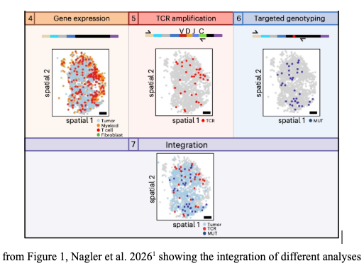
The technology was used to examine mouse model tumors and human patient tumors. Not surprisingly, these analyses revealed that clonally expanded, antigen-specific T cells were spatially colocalized with tumor cells expressing their cognate (neo)antigen². T cells in spatial proximity to tumor cells were found to have diverse functional T cell phenotypes with markers of T cell activation and proliferation but also of T cell exhaustion. T cell phenotypes were influenced by the level of neoantigen expression on the tumor cells and also by the binding strength of the TCR interaction with neoantigen. Strong binding was associated with local enrichment of CD8+ T cells and also of T cell exhaustion signatures, while weak binding was associated with the presence of immunosuppressive macrophages. Sites of productive anti-tumor immune responses could be specifically recognized by signals associated with IFN-γ secretion from activated T cells. Other hallmarks of productive anti-tumor immunity included the presence of dendritic cells and the association of T and B cells into structures called tertiary lymphoid structures (TLS). TLS and the secondary lymphoid organs like lymph nodes are critical sites that support the initial and ongoing anti-tumor immune responses.
A critical element of ongoing anti-tumor immunity is the development of a T cell population that has stem-cell like properties. A second paper from the same team addresses the role of TLS as a source of stem-like progenitor T cells in renal cell carcinoma (RCC)³. The study identified patient samples that contained intratumoural TLS harboring stem-like tumor-specific T cells. In several of these samples they also identified myeloid-rich regions within the tumor where exhausted tumor-specific T cell clones were localized. These descriptions align with classical categories of tumor immune states as ‘cold’ (minimal immune cells present), ‘excluded’ (immune cells blocked from the tumor interior) and ‘hot’ (numerous immune cells present, TLS present). It is notable that in immune-excluded regions within tumors the presence of myeloid cells and cancer-associated fibroblasts (CAF) is commonly observed, with local secretion of TGF-β maintaining immune suppression at the cellular level and CAF-mediated collagen production creating an immune cell barrier.
TLS were then identified as a depot for the stem-cell like T cell population, called in this paper T-progenitor exhausted-like (Tpex). This is a confusing nomenclature that attempts to capture two characteristics – 1) that this cell population has been exposed to chronic antigen stimulation and therefore has features of exhausted T cells (Tex) and 2) that this cell population nonetheless retains self-renewing properties, and are thus stem-cell like ‘progenitors’ (Tp); together: Tpex. Tpex have been found in lymph nodes that are associated with disease sites, eg. tumor-draining lymph nodes⁴. Tpex are a source population for replenishing effector T cells to continue the attack on tumor cells. Notably, the formation of the Tpex population in tumor-draining lymph nodes is dependent on optimal TCR engagement. This echoes themes brought out in the Nagler et al. paper, ie. that optimal TCR binding influences T cell fate in the tumor microenvironment. Linking these two observations is the presence of dendritic cells that provide costimulatory and cytokine support for T cells and present T cells with tumor antigens. Tying all this together then, this second paper now finds Tpex in TLS that are present within the tumor, at least in some RCC patient samples.
These observations generate some interesting hypotheses. First, we can hypothesize that the anti-tumor T cells in the tumor microenvironment and within tumor TLS are decendents of anti-tumor T cells that would have originally been generated in tumor-draining lymph nodes or, alternatively, that anti-tumor T cells arise de novo in the TLS, or that both of these things happen. Second, given the importance of dendritic cells in these systems, we can hypothesize that these cells are mobile and can traffic to lymph nodes or are strictly resident, or again, are both (dendritic cell trafficing is a well understood phenomenon of normal immune responses, but could be subverted in tumors). Finally, that within lymph nodes and within TLS, other signals could undermine productive immune responses even in the presence of T cells and dendritic cells.
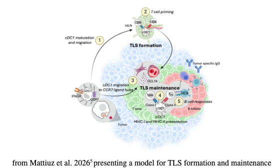
The origin of dendritic cells and T cells in a newly forming TLS was investigated experimentally by Mattiuz et al., writing in Science⁵. Analysis of human cancer samples showed that mature dendritic cells accumulated in TLS, while immature dendritic cells were scattered within the tumor. This suggests that mature dendritic cells, having sensed and acquired tumor antigens, are attracted to nascent TLS. Using a mouse model of lung cancer the process of TLS formation was show to be a multisptep process with initial dendritic cell activation and then migration of dendritic cells to tumor-draining lymph nodes followed by the recruitment of activated T cells to the tumor site and ultimately to TLS. In this model the T cell population arises from the draining lymph nodes, triggered by dendritic cell migration from the tumor. However, in advanced tumors, dendritic cells accumulated directly into TLS with local expansion of T cell effector and Tpex populations.
A preprint paper in bioRxiv tackles the question of T effector cell and Tpex migration, as reported in Nature Reviews Immunology⁶ ⁷. This paper demonstrates in preclinical models that there is a continuous tumor <---> lymph node circuit that sustains stem-like CD8+ T cells ie. the Tpex population. By following intratumoral T cells they showed that effector cells exited the tumor microenvironment and migrated back to the draining lymph node. By doing so these T cells appeared to avoid chronic antigen stimulation that might drive them into exhaustion (Tex). Instead the T cells expressed factors associated with self-renewal and stemness, like TCF1. Antigen presentation within lymph nodes by dendritic cells triggered T cell proliferation and produced a T cell population that could return to the tumor. Two additional observations were striking. First, immune checkpoint blockade with an anti-PD-1 antibody promoted T cell proliferation in the tumor-draining lymph node. Second, the presence of tumor metastases within the tumor-draining lymph node disrupted the Tpex renewal potential, instead promoting terminal exhaustion. As in the RCC “excluded” niche described in Afeyan et al.³, this process was associated with the lack of dendritic cells and the presence of myeloid cells.
Finally, writing in Cancer Cell, Hernández-Verdin et al. identified a factor that contributes to tumor-mediated immune-suppression within TLS⁸. Gamma-aminobutyric acid (GABA) is best known as a brain neurotransmitter but is also synthesized and secreted by some tumor cells, activated B cells and plasma cells. GABA has also been shown to act lymph nodes to dampen inflammation, promote anti-inflammatory/immunosuppressive macrophages, and suppress effector T-cell activity⁹. It seems likely that GABA activity in tumor-draining lymph nodes also has suppressive activity, linking back to the Delclaux et al. paper⁶. Of course, using GABA inhibitors to manipulate anti-tumor immune responses would require novel compounds that do not cross into the blood brain barrier, as the side effects of blocking GABA in the CNS are severe. Since the presence of B cells in TLS and lymph nodes has long been known to support anti-tumor immunity, the signals underlying the switch from anti-tumor B cell friend to GABA-secreting foe are of interest.
Stay tuned.
References
Nagler et al. July 2026; doi: 10.1038/s41587-026-03194-1
A neoantigen is an antigen that is created by the mutations that occur within tumor cells. Neoantigens are never found in normal cells.
Afeyan et al. 22 July 2026 in Nature; doi: 10.1038/s41586-026-10808-w
Lan et al. May 2024 in Nature Immunology; doi: 10.1038/s41590-024-01843-8
Mattiuz et al. 16 July 2026 in Science; doi: 10.1126/science.ady1678
Delclaux et al. May 2026 on bioRxiv; doi: 10.64898/2026.04.30.721705
Yadav & Zamarin. 22 July 2026 in Nature Reviews Immunology; doi: 10.1038/s41577-026- 01342-x
Hernández-Verdin et al. July 2026 in Cancer Cell; doi: 10.1016/j.ccell.2026.06.006
Zhang et al. 3 Nov 2021 in Nature; doi: 10.1038/s41586-021-04082-1